Click image to close
Previous
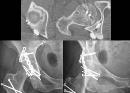
Figure 3 A-C: Postoperative axial CT images through the roof of the acetabulum and hip illustrating anatomical reduction and acceptable positioning of the hardware, and AP and Obturator Oblique radiographic pelvic views (counterclockwise from top) at six months following surgery reveal maintenance of fi xation and joint space.