Click image to close
Previous
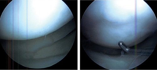
Figure 4. Arthroscopic views of the right knee demonstrate the lateral meniscus to have normal size, position, and morphology with intact horn attachments. The articular cartilage on the osteochondral allograft in both the lateral femoral condyle and the lateral tibial plateau appeared intact. The junction between the osteochondral plug in the lateral femoral condyle and the surrounding native articular cartilage could still be discerned.