Click image to close
Previous
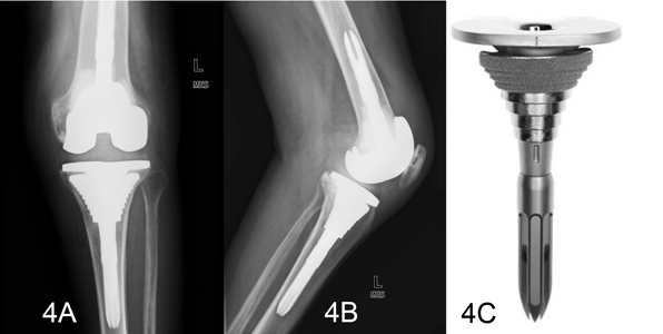
Figure 4. Antero-posterior (4A) and lateral (4B) radiographs two years post-operatively showing proper alignment and fixation of both components. A sample of the tibial sleeves is shown (4C).