Accessory Navicular
Medically reviewed by John S. Blanco, MD ; Emily R. Dodwell, MD, MPH, FRCSC ; Shevaun Mackie Doyle, MD ; David M. Scher, MD
Summary: This article explains accessory navicular is – abnormal, extra bone on the inside of the foot – and common pain symptoms, especially in older children and adolescents. It reviews the different types of accessory navicular, other factors that may trigger discomfort such as flat feet, injury, or overuse. Readers will learn how it is diagnosed, available conservative treatment options, and when surgery may be considered.
On this page:
What is an accessory navicular?
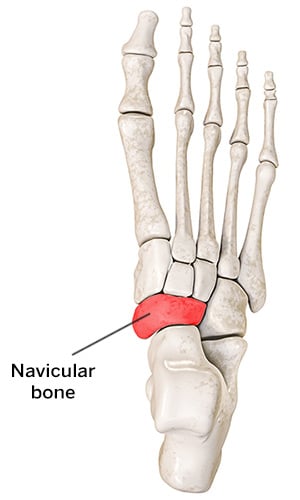
Also known as os naviculare or os tibiale externum, an accessory navicular is an extra bone on the inside of the navicular (the bone in the middle of the arch of the foot) and within the posterior tibial tendon that attaches to the navicular bone.
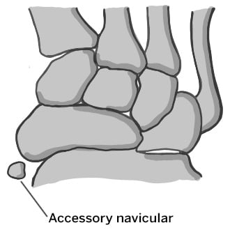
Top-view of accessory navicular in the right foot
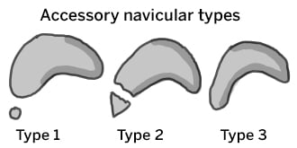
Types of accessory navicular
There are three types of accessory navicular: Type 1 is small, round, and exists within the tibialis posterior tendon. Type 2 is larger and connects to the navicular via a cartilage bridge (called a synchondrosis). Type 3 is the most prominent and connects to the navicular via a bony bridge.
Accessory ossicles or extra bones are common and most often are asymptomatic. There are more than 20 possible accessory bones in the foot and ankle.1
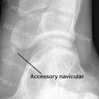
External oblique view X-ray of a type 1 accessory navicular
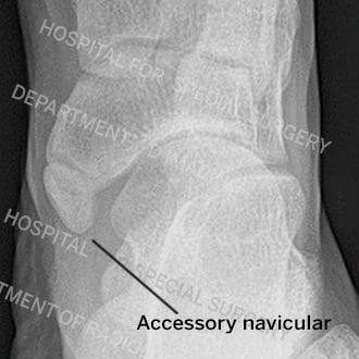
External oblique view X-ray of a type 2 accessory navicular
What are the symptoms of accessory navicular?
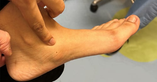
The main symptoms are pain during weightbearing, redness, and localized swelling in the medial (inside) arch of the foot in older children and adolescents. Sometimes, the pain may cause a limp or difficulty with wearing shoes.
How common is accessory navicular syndrome?
Approximately 10% to 14% of normal feet have an accessory navicular.2
Does accessory navicular syndrome get worse with age?
Accessory navicular syndrome does not get worse with age. If it goes untreated, symptoms may linger regardless of an individual’s age.
What causes accessory navicular syndrome?
Ill-fitting shoes, flat feet, foot/ankle sprains, or athletic overuse may cause accessory navicular syndrome. As children mature, the accessory navicular calcifies and becomes firm. This may cause direct pressure on the skin between the shoe and the accessory navicular. Sometimes, repetitive motion or sudden impact across the cartilage bridge between the accessory navicular and the navicular may become inflamed and irritate the middle of the arch causing persistent pain. Additionally, the posterior tibial tendon that attaches to the navicular may become irritated and inflamed.
Is accessory navicular genetic?
Accessory navicular can be transmitted from parent to child as an inherited trait. This has been shown in the literature through studies of families with multiple members who have the condition.3
How do you treat an accessory navicular?
Rest, especially after a sprain or overuse, along with ice, elevation, and over-the-counter anti-inflammatory medication may alleviate accessory navicular syndrome. Modifying shoe wear or inserting soft orthotics to eliminate direct pressure over the middle of the arch of the foot may also help. Physical therapy focused on calf stretching and ankle stabilization may provide additional relief. When all these measures fail, immobilization in a short leg cast or walking boot for 4 to 6 weeks may provide enough rest to the irritated region to eliminate symptoms.
When is surgery needed for accessory navicular?
If non-operative management fails and accessory navicular syndrome persists and/or interferes with everyday activities and participation in sports, the accessory navicular should be removed surgically. This is an outpatient procedure and generally requires four weeks of rest in a cast, splint, or walking boot.
Key takeaways
- An accessory navicular is extra bone tissue in the foot that affects about 10% to 14% of people.
- Most people feel no symptoms, but some (especially teenagers) experience pain, swelling, or difficulty wearing shoes.
- Symptoms may be aggravated in people who have flat feet or poorly fitting shoes, or in those who perform repetitive activity or injure their foot.
- Conservative treatments include rest, ice, anti-inflammatory medications, different footwear, orthotics, and physical therapy.
- If symptoms do not improve with nonsurgical care, outpatient surgery to remove the bone may be recommended.
Explore Related Patient Stories
View All Patient Stories
Owen F
Wyckoff, NJ
Accessory Navicular

Riley Vergano
Long Island, NY
Accessory Navicular
References
- Murphy, Robert F. MD; Van Nortwick, Sara S. MD; Jones, Richard MD; Mooney, James F. III MD. Evaluation and Management of Common Accessory Ossicles of the Foot and Ankle in Children and Adolescents. Journal of the American Academy of Orthopaedic Surgeons 29(7):p e312-e321, April 1 2021.
- Geist, Emil MD. The Accessory Scaphoid Bone. Journal of Bone and Joint Surgery 7(3):p 570-574, July 1925.
- Dobbs MB, Walton T. Autosomal dominant transmission of accessory navicular. Iowa Orthop J. 2004;24:84-5.