Osteochondral Lesion of the Talus
An osteochondral lesion of the talus is a small fracture or lesion of the cartilage that covers the talus, which is the lowest bone of the ankle joint. In some cases, there may also be damage to the bone itself. This condition is sometimes called an osteochondral fracture or osteochondritis dissecans. However, it should not be confused with osteochondritis dissecans of the knee.
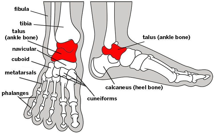
Illustration of the bones of the foot and ankle shown from the top and side view, with the talus highlighted.
What causes an osteochondral lesion of the talus?
This condition may be caused by either a traumatic injury, such as a sprained ankle, or over time to repetitive movement or overuse. The majority of OLTs occur after a traumatic ankle injury or ankle sprain. Repetitive ankle sprains and chronic ankle instability can also result in OLT. Approximately 27,000 people experience ankle sprains each day in the U.S. and OLTs have an incidence of between 50% and 70% of all acute ankle sprains.
What are the symptoms of an osteochondral lesion of the talus?
The main symptom is ankle pain, often when standing or weight bearing. As cartilage injuries have a poor spontaneous healing response, symptomatic OLTs often require surgical treatment.
How is an osteochondral lesion of the talus diagnosed?
Foot and ankle surgeons may have a suspicion for OLT from a patient’s history and symptoms. Routine x-rays often miss up to 50% of these injuries as it is unable to detect overlying cartilage injuries. Therefore, an MRI is recommended for a definitive diagnosis with high sensitivity to cartilage and bone changes. MRI can also detect other related pathology, such as ligament or tendon injuries.
How is an osteochondral lesion of the talus treated?
Lesion size and location determine the appropriate treatment strategy. Reparative procedures such as bone marrow stimulation is indicated for lesions of less than 10 mm in diameter and performed by minimally invasive arthroscopic techniques. Injured cartilage and bone are removed to promote healing stimulation. Larger lesions are best treated with replacement procedures including autologous osteochondral transplantation.
Biological agents such as platelet-rich plasma (PRP) and bone marrow aspirate concentrate (BMAC) have been more recently studied as an adjunct to surgical treatment and good evidence of effectiveness is established. These biologics release a number of cytokines and growth factors that stimulate the healing of cartilage and bone. OLTs are common. Any player who has an ankle sprain that is not resolving should consider an MRI to out rule these cartilage injuries. Treatment, when started soon, shows excellent outcomes with the addition of biologic adjuvants. These outcomes have been improving.
Osteochondral Lesion of the Talus Success Stories
In the news
Updated: 12/6/2016