Femur Fractures in Children: Treating a Child’s Broken Thighbone
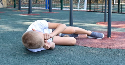
Despite the size and strength of the femur (the thighbone), femoral fractures in children are common. A motor vehicle accident, a fall from a piece of playground equipment, or even a piece of furniture at home may result in a broken leg that can range in severity from a simple hairline crack to a complex injury that damages surrounding soft tissues.
What are the treatments for a broken femur in children?
As in an adult, the treatment goals for a child with a broken femur include achieving proper realignment of the bone, promoting rapid healing, and returning the patient to normal activities. However, there are also unique factors and considerations in the treatment of broken bones in children. These factors include:
- The configuration of the fracture. The most common patterns are:
- Transverse fracture (straight across the shaft of the bone).
- Oblique fracture (a fracture across the bone at an angle).
- Spiral fracture (a longer fracture that usually spans a longer segment of the bone).
- The "energy" of the injury.
- The amount of soft tissue injury present.
- The patient’s size and age.
- The amount of growth the child has remaining.
- The potential for any shortening of the bone that can sometimes occur with a fracture of a growing bone.
How are femoral fractures in newborns and infants treated?
Femur fractures in newborns are unusual. However, they may occur, for example, in babies born with a skeletal dysplasia such as osteogenesis imperfecta − also known as brittle bone disease − or sometimes following a difficult delivery. Fractures in these children and in infants are usually treated by placing the child in a Pavlik harness, a cloth brace that helps hold the thigh in the proper position during recovery while it heals.

Pavlik harness on a child with developmental hip dysplasia.
At this young age, children’s bones heal very rapidly, usually by three to four weeks.
Unlike the healing process in adults, a certain amount of variation in alignment of the bone as it heals is acceptable in infants and older children. As the body lays down new bone, over time, there is an automatic "correction" or straightening during growth, called remodeling.
How are toddlers and young children treated for fractures of the femur?
Among children ranging from toddlers up to five, femur fractures usually result from a low energy fall, and a special type of cast called a spica cast is the most common treatment. In most cases, the orthopedic surgeon realigns the fracture using fluoroscopy or X-ray imaging as a guide and immobilizes the leg in the spica cast. This procedure usually takes place in the operating room and a pediatric anesthesiologist is present to administer a sedative or general anesthesia to keep the child comfortable.
While casting techniques vary among orthopedic surgeons, for femur fractures, the spica cast usually extends from mid-chest down the length of the affected leg and halfway down the other leg.
This positioning helps maintain alignment of the fracture and reduces the risk of it moving out of place. Some children have fractures that are stable enough to treat them in a walking spica cast. This cast extends from the mid chest to the length of the injured leg, but leaves the uninjured leg free to facilitate walking.


A young patient in a spica cast, which immobilizes the leg after realignment.
Children remain in the spica cast for a period ranging from four to eight weeks, but sometimes as long as three months, which can be challenge for caregivers when it comes to maintaining hygiene and keeping the child distracted and happy. Though cumbersome, spica casting continues to be the continues to be the safest and least complicated method for treating fractures among this age group and yields excellent results.
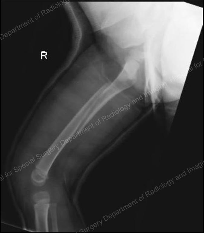
X-ray of a femur fracture with spica cast.
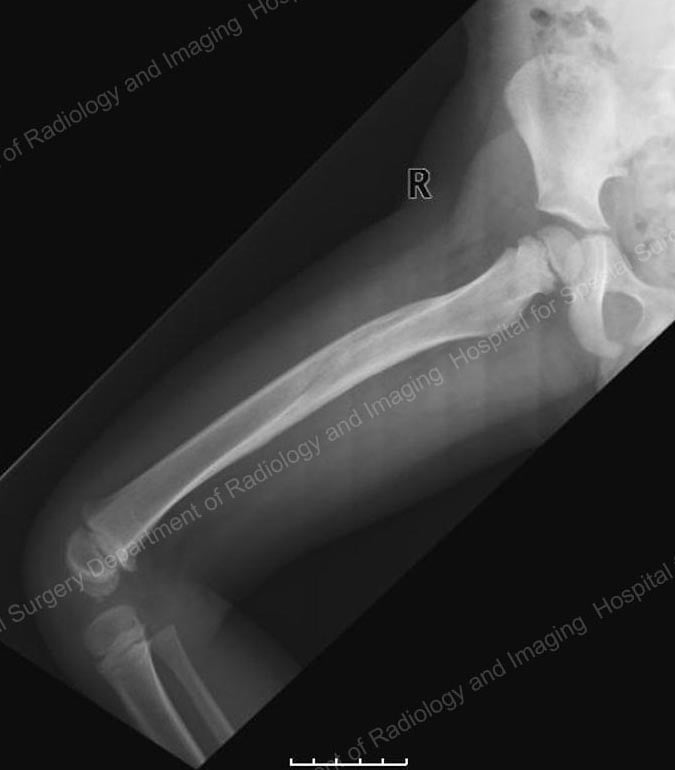
X-ray 10 months after injury and removal of spica cast.
Following treatment, the orthopedic surgeon continues to monitor the patient for a period of several years to ensure that there is no limb length discrepancy. When a fracture in the shaft of the femur occurs, the bone tends to shorten at the point where it breaks. Later on, as healing occurs among children in this age group, the bone tends to grow longer; this is a phenomenon called "overgrowth" that may be caused by increased blood flow to the fracture site.
In some patients this initial shortening, combined with the subsequent increased growth, "cancel" one another out. However, in those cases where a small limb length discrepancy occurs, the orthopedic surgeon can use a relatively simple technique, such as growth modulation of the longer leg, to address this issue.
Treatment for Children aged 5 to 10 years
Intramedullary nails or rods
Since the early 2000s, many pediatric orthopedists have started using intramedullary nails or rods made from strong, lightweight, and flexible titanium to stabilize femur fractures in children aged five and older. In a relatively simple technique, the orthopedic surgeon makes two small incisions − about one inch in length − on either side of the knee. After the bone is realigned, the nails are inserted up through the center of the bone where they act as an internal splint during healing.
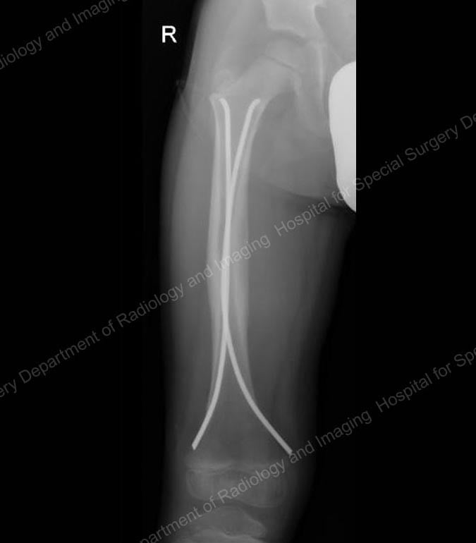
X-ray showing flexible intramedullary nails in place.
Intramedullary nails come in a range of diameters to accommodate the varying size of children’s bones. Generally no casting is necessary, however a knee immobilizer is commonly used for a few weeks to prevent movement and to keep the child comfortable. In most cases, a recovery period of three to six weeks of early healing is necessary before the child can begin walking on the injured leg. When the bone is completely healed, usually around one year after the injury occurs, the child returns to the hospital to have the nails removed.
Intramedullary nails are usually very well-tolerated. They are particularly useful in five- to ten-year-old children who have a thick lining around the bone called the periosteum, which might be likened to the peel on a banana. The periosteum helps keep the bone more stable and reduces the need for a heavy and rigid device, such as those that might be used in adults.
Treatment for older children and adolescents
Intramedullary nailing can also work well in older or heavier children, typically those over age 10 and heavier than 100 pounds. However, a single, thicker and more rigid nail is used rather than the smaller flexible nails.
In these children and teenagers, the nail is inserted through the top of the femur, near the hip, at a point called the greater trochanter, and locked into place with a screw at the top and the bottom of the bone. This placement avoids interruption of blood flow to the ball of the hip joint, which can lead to disabling arthritis. These concerns are not present in adults because they have different anatomy. In order to minimize the risk of complications when using intramedullary nailing in children, the orthopedist uses different devices and techniques than they would use in adult patients.
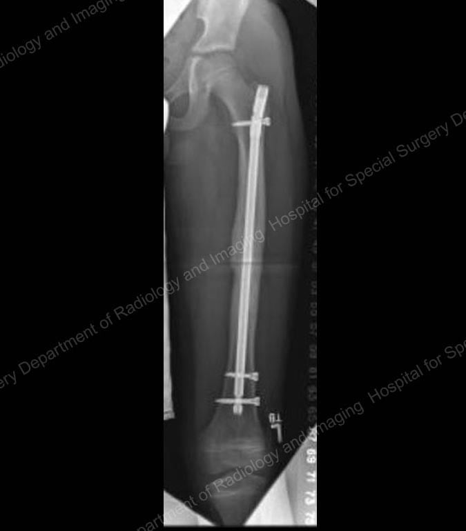
X-ray showing a rigid intramedullary nail in place.
Submuscular plating
Submuscular plating is another treatment option for older and larger children. In this procedure, using X-ray images for guidance, the orthopedic surgeon inserts a stainless-steel plate under the muscles of the leg and across the fracture site. Through small incisions in the skin, screws are placed through the plate and bone, above and below the fracture to hold it in place. Although this technique is minimally invasive, removal of the plate is more difficult than removal of intramedullary nails. Currently, there is some discussion ongoing in the orthopedic community regarding the need to remove the plates at all.
External fixation
In addition to the approaches described, the fallback treatment for a femur fracture in a child of any age is external fixation.
This technique involves the use of rigid metal rods or circular rings and pins inserted into the bone to maintain alignment during healing.

An external fixation ("ex-fix").
How has treatment for femoral fractures evolved?
The use of intramedullary nails, external fixators, and other stabilization devices represent a dramatic improvement over the traditional approach to fractures of the femur. As recently as the 1980s, the standard approach for most children over the age of two years was placement of a pin through the bone just above the knee and traction; this typically involved a hospital stay of three weeks or longer in which many adjustments were needed to maintain alignment. Traction was followed by placement in a body cast for as long as three months.
While this method resulted in excellent healing, proper alignment, and strong bones, it also required significant involvement on the part of a parent or other caregiver. Fortunately, the available technology and our surgical skills have advanced to a point where we can achieve these same results in a more time-efficient manner which is much less disruptive to the family. Today we are able to make our treatment decisions based not only on how we can best get the fracture to heal, but also on choosing the method that will be best tolerated by the child and his or her family.


