Instrumented Cervical Laminectomy, Fusion
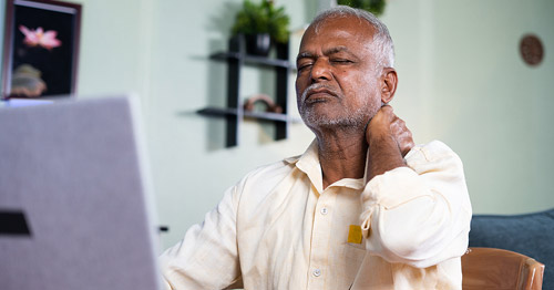
Decompression neck surgery for patients with spinal stenosis
A cervical laminectomy removes small bony structures (the spinous process and lamina) from one or more vertebrae of the neck region of the spine to alleviate pressure on the spinal cord, which causes pain. This is a common treatment for cervical spinal stenosis, a condition in which there is a narrowing of the spinal canal.
Spine anatomy
The spine, or backbone, is the center of support for the upper body. This column of bones and cartilage extends from the base of our skull to the pelvis, enclosing and protecting the spinal cord. Ligaments and tendons and large muscles connect to the spine, while highly sensitive nerves extend outward from the spinal cord. The spinal cord extends from the base of the brain and enters the spinal canal, a protective space within our bony spine, to carry signals to and from the brain throughout the body.
A healthy spine is both highly flexible and very strong. Its strength holds up our head and shoulders and supports our upper body. It allows us to stand up straight. The flexibility of the spine enables us to bend and twist.
Vertebrae
The spine is not one long rigid bone. It consists of 24 small bones called vertebrae that are stacked in a column from the pelvis to the base of our skull. These bones connect to create a canal that protects the spinal cord.
The vertebrae are divided into four regions. From top to bottom, these are:
- The cervical spine (the neck) – the first seven vertebrae located just below the skull
- The thoracic spine – the 12 vertebrae of the upper back
- The lumbar spine – the five vertebrae of the lower back
- The sacral spine – composed of a triangular structure called the sacrum (five individual vertebrae that fuse together between the age of 18 and 30) and the coccyx (commonly called the tailbone and composed of three to five individual vertebrae, some of which may fuse together in adulthood)
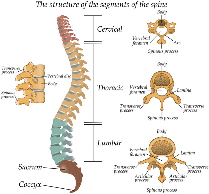
Lateral (side) view of the spine in a person facing to the viewer's left.
The curves of the spine
The spine is not perfectly straight; it has natural curves. If you were looking at the spine from the side, you would see that it is curved like an elongated 'S'. These natural curves are very important. When properly maintained, they give the spine full mobility and provide stability for the backbone and surrounding trunk. Good posture is important to maintain the health of our spine.
Spinal cord and nerves
The spinal cord is a cylinder of nerve tissue. Roughly the thickness of a finger, it extends from the skull to the lower back, traveling through the middle part of each stacked vertebra, called the central canal. Nerves branch out from the spinal cord through openings in the vertebrae and carry messages between the brain and the muscles.
Spinal discs
Structures called intervertebral discs are located between each vertebrae. They are flat and round, and about half an inch thick. Their main purpose is to provide shock absorption and allow mobility between the vertebrae. The structure of a spinal disc can be likened to that of a jelly doughnut. It has an outer ring of firm, spongy, malleable material and an inner core composed of a jelly-like substance.
A herniated disc is a common condition that affects these soft tissues. A disc herniation (sometimes called a "slipped disc") occurs when a portion of a disc's inner core bulges or ruptures through the outer ring, putting pressure on a spinal nerve. This may cause low back pain and/or leg pain – such as that associated with sciatica – leg weakness, leg numbness, cauda equina syndrome and other problems.
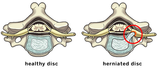
Illustrations of the axial (overhead) view of a healthy disc and a herniated disc putting pressure on a spinal nerve.
Spinal musculature
The spine is supported and controlled by several layers of muscles that perform different actions, yet work together in a harmonious fashion to support the spine, hold the body upright and allow the trunk to move, twist and bend.
Long and thick muscles span much of the back and function like guide wires, protecting the spine from excessive and sudden movement.
Deep and thinner muscles connect from the rib cage to the pelvis and hips. Together, these muscle groups act as a natural corset to provide stability and a foundation from which the hips and pelvis can derive power. This is known as core musculature.
Animation: Instrumented cervical laminectomy with fusion
This animation provides an inside look into an instrumented cervical laminectomy with fusion. In this procedure, a laminectomy of the cervical spine is performed to release pressure on the spinal cord. Instrumentation and, in some cases, bone grafts are added to fuse the vertebrae and stabilize the spine where bone was removed.
How instrumented cervical laminectomy with fusion is performed
Incision and removal
An incision is made along the midline at the back of the neck. After the spine is exposed, surgical instruments are used to remove the spinous process and lamina from the affected vertebrae. Another variation of this step involves using a motorized instrument to cut a trough through the lamina on both sides of the vertebra and removing the lamina and spinous process as a single piece. A surgical microscope is often used during this procedure to ensure a high degree of accuracy.
Preparing for fusion
A motorized instrument (bur) is used to remove the top (cortical) layer of the articular processes (spine joints) at the sides of the vertebrae to prepare a site for fusing the vertebrae. The fusion will stabilize the spine where bone was removed.
Graft placement
Bone grafting can be done with pieces of a patient’s own bone (autograft), processed bone from a bone bank (allograft), or a bone graft substitute (demineralized bone, ceramic extender, or bone morphogenetic protein). When a patient’s own bone is used for grafting, the pieces are typically crafted from the bone components that were removed previously (local bone graft). Bone grafts are placed along the prepared site where the top layer of bone was removed. This bone eventually grows in place, fusing the spine and providing additional stability.
Closure and recovery after cervical laminectomy
The incision is closed and dressed to complete the procedure. A cervical collar is often worn for six weeks following surgery. Patients should be careful to avoid heavy lifting and excessive neck motion during recovery.
Updated: 4/26/2019