Spine Surgery: Thoracic Laminectomy With Instrumentation, Fusion
Decompression back surgery for relief of spinal stenosis and tumor removal
Spinal stenosis is a narrowing of the spinal canal, which can cause painful pressure on the spinal cord or nerves. Sometimes the source of this narrowing is a tumor that has spread to the thoracic region of the spine and is pushing on the spinal cord.
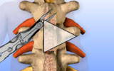
A thoracic laminectomy removes the lamina portion of vertebrae to provide access to the tumor to remove it and eliminate pressure on the spinal cord. After removing bone, instrumentation can be added to stabilize the vertebrae.

View thoracic laminectomy animation
Incision and removal
An incision is made along the middle of the back. Once the spine is exposed, surgical instruments are used to remove the spinous processes. Next, the lamina portions of affected vertebrae are removed, providing access to
Tumor removal
Surgical instruments are used to remove the tumor. Removing both the tumor and overlying bone that was pushing it into the spinal cord relieves the compression and pain.
Stabilizing the spine
Instrumentation is introduced to support the spine. Holes are made in the pedicle of intact vertebrae and screws are placed in the drilled holes. Next, rods are positioned between the screws and fastened in place. The rod and screw instrumentation provides stability to the spine.
Closure and recovery
The incision is closed and dressed to complete the surgery. Radiation treatments are frequently used two to four weeks after surgery to treat any tumor remaining in the spine. Adding the instrumentation after the laminectomy increases the strength of the spine and may decrease the need for a post-operative brace. Patients should avoid heavy lifting, bending, twisting, and turning for six to twelve weeks.