Lisfranc Fracture Dislocations
Case Example
A 48 year-old male was involved in a high speed car accident and sustained a mid-foot injury. Radiographs revealed a Lisfranc fracture-dislocation. He was referred to Dr. David L. Helfet at the Orthopedic Trauma Service of Hospital for Special Surgery for definitive management. The Lisfranc fracture-dislocation was reduced and fixed through a minimally invasive technique with placement of multiple screws. He most recently returned for routine follow-up at 3 months and radiographs illustrate a healing Lisfranc fracture dislocation and he has resumed all activities of daily living with significant improvement of pain and swelling.
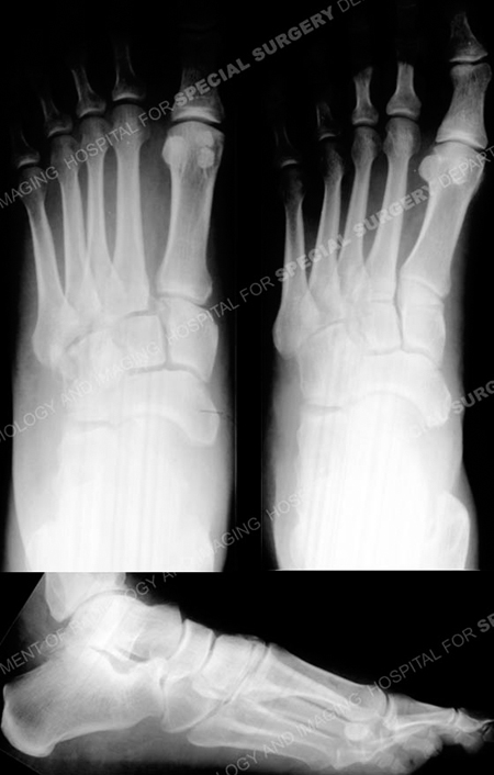
Anteroposterior, oblique, and lateral radiographs revealing a Lisfranc fracture-dislocation.
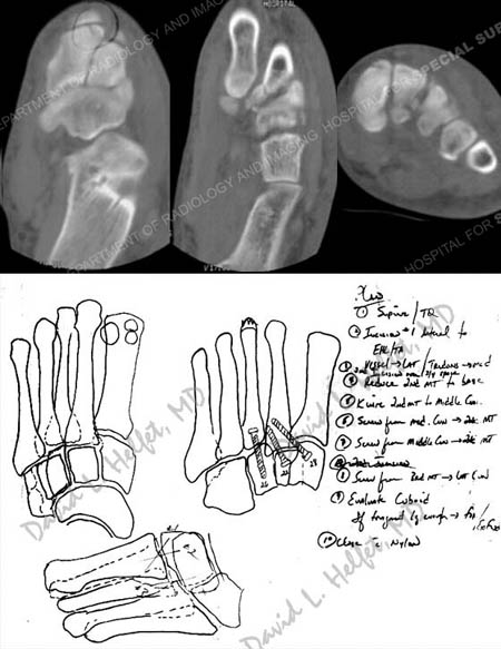
CT scan images further delineating the fracture pattern and pre-operative plan.
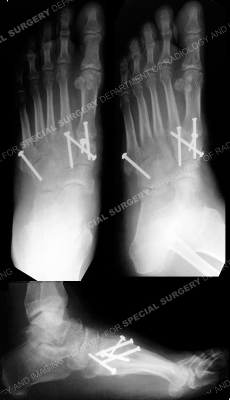
Anteroposterior, oblique, and lateral radiographs at 3 months illustrating a healing Lisfranc fracture-dislocation.
Research Publications
The HSS Orthopedic Trauma Service has conducted many studies. Please see our publications on treatment of fracture dislocations, foot fractures, and minimally invasive fracture treatment.