Pathologic Fractures
Case Example
A 70-year-old male slipped and fell while skiing, landing on his outstretched right arm. Radiographs at the local hospital revealed a right-sided pathologic mid-shaft humeral fracture and evidence of lytic lesions on the humeral head. His right arm was placed in a fracture splint and he was referred to David L. Helfet, MD at the Orthopedic Trauma Service at Hospital for Special Surgery for definitive fracture management. Additional tests were performed to further evaluate the pathologic fracture and metastatic lytic lesions. Fracture reduction and fixation was planned and performed using an expandable retrograde intramedullary humeral nail. He followed up with an oncologist for further evaluation and treatment. He returned for regular follow-up and his pathologic humeral fracture healed uneventfully. At his latest follow-up appointment at 6 months he presents with excellent radiographic and clinical results including a healed pathologic humerus fracture in excellent alignment with maintenance of reduction and fixation and he returned to his activities of daily living with resolution of pain.
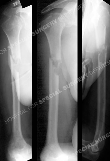
Anteroposterior and lateral radiographs reveal a right-sided pathologic mid-shaft humeral fracture and evidence of lytic lesions on the humeral head.
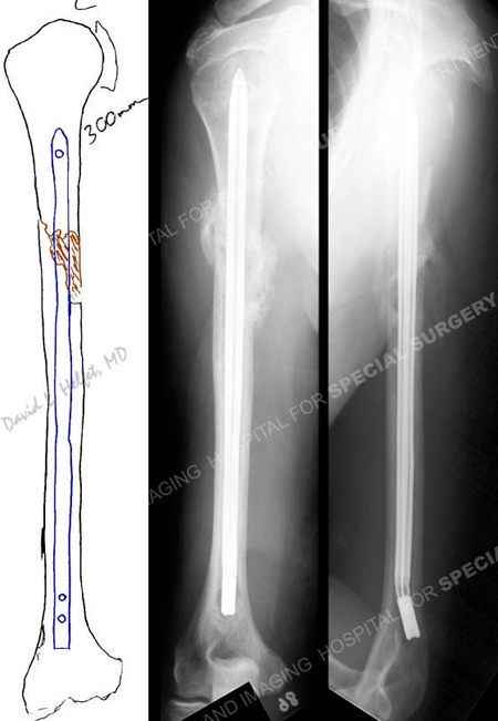
Pre-operative plan and anteroposterior and lateral radiographs at 6 months illustrating a healed mid-shaft pathologic humeral fracture in excellent alignment.
Research Publication
The HSS Orthopedic Trauma Service has conducted many studies. Please see our publications on pathologic fractures, fracture treatment in older populations, and humerus fractures.