Polytrauma
Case Example
A 48-year-old male was involved in a motorcycle accident, after striking a deer which had entered the roadway. The patient was brought to our emergency department and radiographs revealed a left-sided pelvic fracture with a sacral and superior and inferior pubic rami fractures and a right-sided calcaneus fracture. He was placed under the care of David L. Helfet, MD at the Orthopedic Trauma Service of Hospital for Special Surgery. Anterior external fixation was placed for initial stabilization of the pelvic fracture. Open Reduction and Internal Fixation (ORIF) was then performed for the calcaneus fracture with placement of with placement of a plate and screws and supplemental bone graft. Three days later, definitive surgery was performed for the pelvis fracture with reduction of the sacral fracture and placement of a percutaneous screw across the sacroiliac (SI) joint stabilizing the sacral fracture, and the pubic rami fractures were reduced and stabilized with a pelvic reconstruction plate and screws. He most recently returned for routine follow-up at 6 months following surgery and radiographs revealed healed pelvic and calcaneus fractures and he had fully resumed his occupation and all activities of daily living.
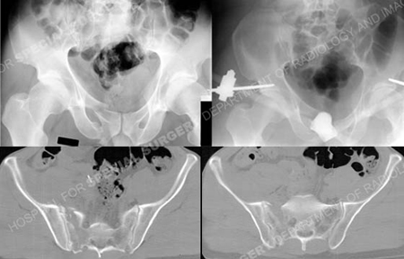
Anteroposterior injury pelvic radiograph (top left image) revealing a left-sided pelvic fracture including a sacral fracture and superior and inferior pubic rami fractures, anteroposterior pelvic radiograph following initial closed reduction and placement of anterior pelvic external fixation (top right image), and CT scan images further illustrating the sacral fracture pattern.
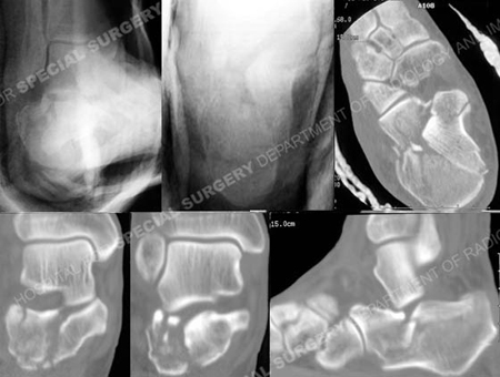
Plain radiographs and CT scan images of the right foot revealing a right-sided calcaneus fracture.
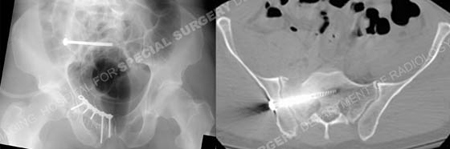
Postoperative anteroposterior pelvic radiograph and CT scan image illustrating acceptable reduction and placement of hardware.
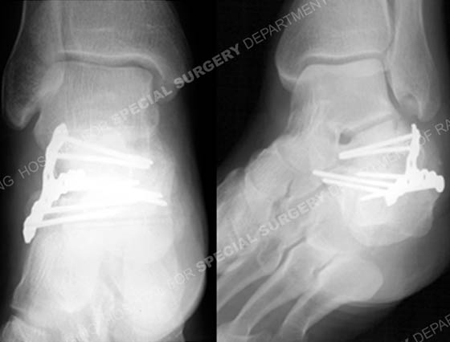
Postoperative radiographs at 6 months demonstrating a healed calcaneus fracture.
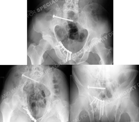
Postoperative radiographs at 6 months demonstrating healed pelvic fractures.
Research Publications
The HSS Orthopedic Trauma Service has conducted many studies. Please see our publications on pelvic fractures.