Ulna Fractures
Case Example
A 76-year-old female slipped and fell onto her left upper extremity and elbow. She was taken to a local hospital and radiographs revealed a closed left-sided ulna olecranon fracture. She was referred to David L. Helfet, MD at the Orthopedic Trauma Service of Hospital for Special Surgery for definitive management. Open Reduction and Internal Fixation (ORIF) was performed and the fracture was reduced and fixed using a tension band construct. She returned for regular follow-up and healed uneventfully. The hardware was removed 7 months following surgery. At the time of her latest follow-up visit, 10 months following fracture surgery, she has excellent radiographic and clinical results including a healed ulna fracture in excellent alignment, resolution of pain, and a return to pre-injury activities.
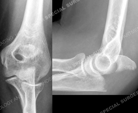
Anteroposterior and lateral radiographs revealing an ulna olecranon fracture.
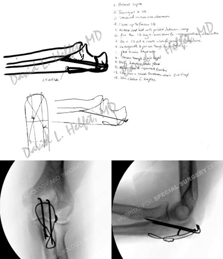
Pre-operative plan (top image) and intraoperative fluoroscopic radiographs (bottom images) following ORIF.
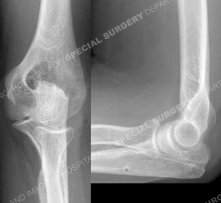
Anteroposterior and lateral radiographs 10 months following fracture surgery illustrating a
healed olecranon fracture in excellent alignment.
Research Publications
The HSS Orthopedic Trauma Service has conducted many studies. Please see our publications on ulna fractures and fracture treatment in older populations.