Correction of Post-Traumatic Deformity
Case Example
Mid-shaft Tibial Nonunion with Deformity
A 49 year-old male sustained a right-sided open mid-shaft tibia fracture after being struck by a stolen pickup truck while riding his bicycle. He was treated at an outside hospital with irrigation and debridement and insertion of an intramedullary (IM) nail. He continued to follow-up at the treating hospital and developed a draining wound near the fracture site and was diagnosed with an MRSA infection and treated by intravenous antibiotics. The intramedullary nail was eventually removed and several attempts at non-operative management were attempted at the outside hospital and he was told that his infection was eradicated. He came to see Dr. David L. Helfet at the HSS Orthopedic Trauma Service one year after his injury for a second opinion (his antibiotic treatment had been discontinued 6 months prior). Radiographs were again obtained and revealed a mid-shaft tibial nonunion with significant deformity of approximately 30° varus and 15° flexion. Correction of deformity was planned and performed with debridement of the nonunion site and insertion of a reamed tibial nail with a proximal dynamic interlocking screw. The patient returned for regular follow-up visits and radiographs at 6 months post-operative illustrate a healed tibia nonunion and he reported a return to pre-injury activities and resolution of pain. The IM nail was removed at 1 year following the index surgery.
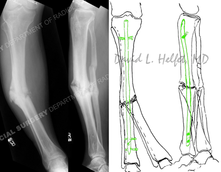
Radiographs (left images) illustrate a right-sided mid-shaft tibial nonunion with 30° varus deformity and 15° flexion deformity; pre-operative (right image) plan for correction of deformity and insertion of a reamed intramedullary (IM) nail.
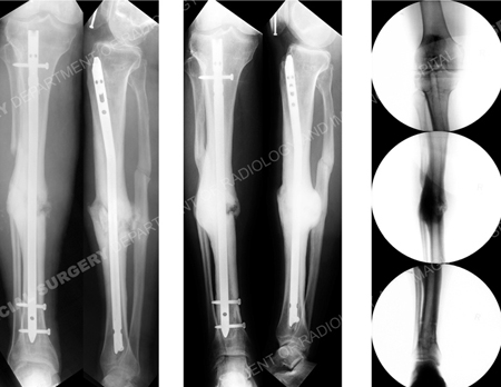
Immediate postoperative radiographs (2 left images) following correction of deformity and insertion of a reamed IM nail and screws including a dynamic proximal dynamic interlocking screw; radiographs 1 year following the index surgery (middle 2 images) illustrate a healed tibia nonunion; fluoroscopic images (right image sequence; top to bottom) following removal of the reamed IM nail (13 months following the index surgery).
Research Publications
The HSS Orthopedic Trauma Service has conducted many studies. Please see our publications on tibia fractures, open fractures, deformity correction, and nonunions.