Fractures in Adolescents
Case Example
Pediatric Acetabular Fracture
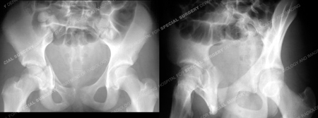
A 13-year-old girl fell while skiing and was taken to a local hospital. Radiographs were obtained and revealed a right-sided Anterior Column with Posterior Hemi Transverse acetabular fracture. She was placed in skeletal traction and transferred to the care of
Dr. David L. Helfet at Hospital for Special Surgery's Orthopedic Trauma Service for definitive management of her acetabular fracture. Open reduction and internal fixation was performed with placement of contoured plates and screws. She returned for regular follow-up visits and healed uneventfully and at her most recent follow-up visit at 8.5 years following surgery she has excellent clinical and radiographic results and reported complete resolution of pain with full range of motion in the hip joint.

X-rays reveal a right-sided Anterior Column with Posterior Hemi Transverse acetabular fracture.

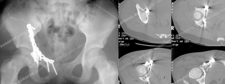
CT scans images further delineate the fracture pattern.

3 dimensional CT reconstructions

Fracture surgery was performed using an ilioinguinal approach. The fracture was reduced and stabilized using a contoured 10-hole pelvic reconstruction plate and screws and placement of a spring plate along the anterior wall fracture. Postoperative CT scan images (right images) illustrate an adequate reduction.

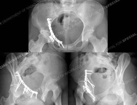
The patient returned at 8.5 years following surgery and radiographs demonstrate a healed acetabular fracture and maintenance of joint space. She had fully returned to full activities and reported no pain.
Research Publications
The HSS Orthopedic Trauma Service has conducted many studies. Please see our publications fractures in adolescents, acetabular fractures, and fractures in athletes.
