Limb Lengthening
Case Example
Acute Limb Lengthening over an Intramedullary Nail
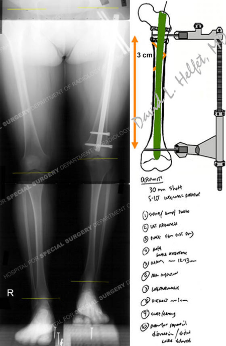
A previously healthy 39 year-old female fell while rollerblading and sustained a left-sided subtrochanteric femur fracture. She was taken to a local hospital and treated with a femoral intramedullary (IM) nail with proximal and distal interlocking screws. She was noted to have shortening postoperatively. She was referred to Dr. David L. Helfet at the Orthopedic Trauma Service at Hospital for Special Surgery at 6 months following surgery for further evaluation and a possible limb lengthening/distraction osteogenesis procedure. Radiographs were obtained and illustrated a healing subtrochanteric fracture in good alignment but with 3 centimeters of shortening. The neurovascular examination was normal and she had full range of motion (ROM) in the knee and hip joints. Her chief complaints were the shortening of her left lower extremity and inability to resume her previous recreational activities.
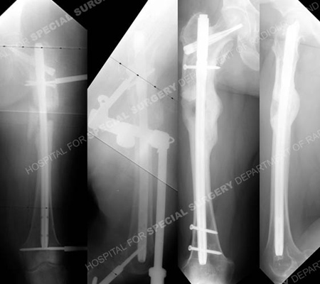
Limb lengthening was planned and accomplished through an acute lengthening procedure with the use of a femoral IM nail and medial butterfly osteotomy, with gradual, in-hospital distraction. Advantages of this technique include the ability to correct deformity in a rapid period of time of 4 days while monitoring the soft tissues and neurovascular status. This time span of 4 days is significantly shorter than the 3-6 months typically associated with conventional limb lengthening procedures. It also avoids the need for extended use of a bulky Ilizarov frame and its associated complications.
Her hospital course was uncomplicated and she was discharged from the hospital 2 days following the procedure. She returned for regular follow-up and fracture union was noted at 4 months postoperatively with complete deformity correction. She last returned for follow-up at 1 year following limb lengthening with excellent radiographic and clinical results including maintenance of fixation, bone union, restoration of limb length, complete resolution of pain, full ROM in the knee and hip joints, and return to pre-injury activities.

Full standing anteroposterior x-ray taken upon initial visit to HSS at 6 months following her injury revealing a left-sided healed subtrochanteric fracture with a limb length discrepancy and 3 cm of shortening on the affected side and pre-operative surgical plan for acute limb lengthening over a femoral IM nail.

Anteroposterior and lateral radiographs (left images) immediately following the osteotomy and exchange nailing with placement of a femoral distractor for the acute limb lengthening procedure. Gradual distraction was performed over the course of 4 days with clinical monitoring and the distal interlocking screws were placed in the IM nail. The limb lengthening procedure was then complete and the femoral distractor was removed. Anteroposterior and lateral radiographs 12 months following limb lengthening (right images) illustrating a healed osteotomy in excellent alignment. Her limb lengths were equal, she reported complete pain resolution and she returned to all pre-injury recreational activities.
Research Publications
The HSS Orthopedic Trauma Service has conducted many studies. Please see our publications on limb lengthening.
