Malunions
Case Example
Mid-Shaft Tibia Stress Fracture
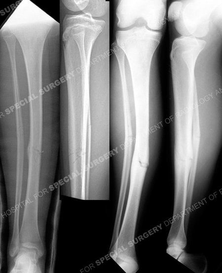
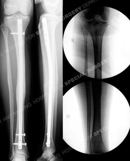
A 15-year-old female, avid soccer and volleyball player, noted progressive onset of right-sided shin pain during her athletic training over the course of several weeks. Radiographs taken at a local hospital revealed a minimally displaced mid-shaft tibia stress fracture. She was initially treated with cast immobilization for 3 months. She was referred to Dr. David L. Helfet at the Orthopedic Trauma Service at Hospital for Special Surgery at 6 months for definitive management of a malunion with bridging callus on the lateral cortex and with 13° of valgus deformity. Operative treatment was planned and performed including correction of the deformity and insertion of an intramedullary (IM) nail and locking screws. She returned for regular follow-up visits and at 3 months following surgery she had excellent radiographic and clinical results including a healed tibia fracture, resolution of pain and full range of motion of the knee and ankle joints. She returned to all pre-injury activities at 4 months. At 1 year following surgery the hardware was removed.

Anteroposterior (AP) and lateral radiographs (left images) reveal a minimally displaced mid-shaft tibia stress fracture. AP and lateral radiographs at 6 months following the injury (right images) illustrating a malunion with 13° of valgus deformity.

Anteroposterior and lateral radiographs 6 months following surgery (left images) illustrating a healed tibial malunion in acceptable alignment. Intraoperative fluoroscopic images (right images) following removal of hardware at 12 months.
Research Publications
The HSS Orthopedic Trauma Service has conducted many studies. Please see our publications malunions, stress fractures, fractures in adolescents, and tibia fractures.
