Periprosthetic Femur Fractures
Case Example
A 66-year-old female tripped and fell while getting out of a car. Her past surgical history included a left Total Knee Replacement (TKR) performed six months earlier. Radiographs revealed a left-sided comminuted periprosthetic femur fracture proximal to the femoral component. A CT Scan was performed and the femoral component was determined to be stable. Through a minimally invasive technique with indirect reduction, the fracture was reduced and fixed using a LISS locking plate and screws. She returned for regular follow-up and healed uneventfully and at 2 years she presents with an excellent outcome including a healed periprosthetic femur fracture, resolution of pain, and a return to pre-injury activities.
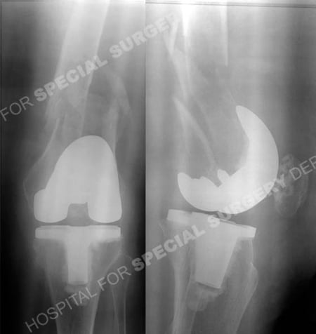
Anteroposterior and lateral injury radiographs revealing a left-sided comminuted periprosthetic femur fracture.
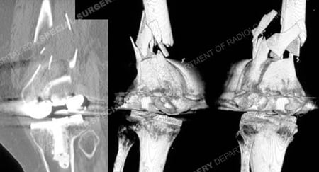
CT scan images and 3D CT reconstruction images further delineating the fracture pattern.
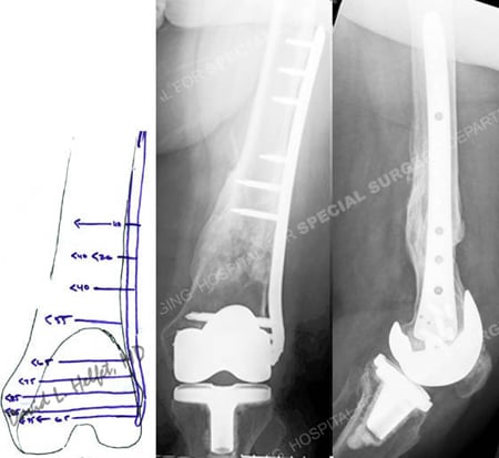
Pre-operative plan (left image) for fracture reduction and fixation with a LISS locking plate and anteroposterior and lateral views at 2 years (right images) following fracture surgery illustrate a healed periprosthetic femur fracture.
Research Publications
The HSS Orthopedic Trauma Service has conducted many studies. Please see our publications on periprosthetic fractures and use of locking plates in fracture treatment.