Wrist Fractures
Case Example
A 65-year-old male was a restrained driver of an automobile struck by another vehicle in a head-on collision while on vacation overseas. He sustained a left-sided wrist fracture and was taken to a local hospital for initial care. Radiographs revealed a displaced distal radius fracture with articular involvement. Spanning external fixation was placed at the outside hospital for initial management. He was referred to David L. Helfet, MD at the Orthopedic Trauma Service of Hospital for Special Surgery for definitive management of his wrist fracture. Open reduction and internal fixation was performed with placement of a locking plate dorsally. He returned for regular follow-up and radiographs at 6 months illustrate a healed distal radius fracture in anatomic alignment with maintenance of hardware and he reports resolution of pain and a return to pre-injury activities.

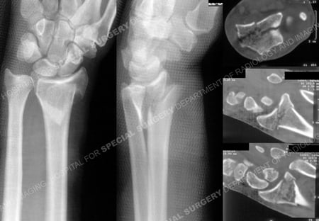
Anteroposterior and lateral radiographs (left images) illustrating a left-sided displaced distal radius fracture with articular involvement and CT scan images (right images) further delineate the fracture pattern
and articular involvement.

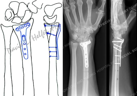
Pre-operative plan for open reduction and internal fixation with a dorsal locking plate and screws (left image) and radiographs at 6 months (right images) reveal a healed distal radius fracture.
Research Publications
The HSS Orthopedic Trauma Service has conducted many studies. Please see our publications on wrist fractures, use of locking plates in fracture treatment, and radius fractures.
