Femoral Anteversion (Hip Anteversion)
In anatomy, the word "version" refers to the angle or rotation of all or part of an organ, bone or other structure in the body, relative to other structures in the body. Anteversion refers to an abnormal forward rotation.
- What is femoral anteversion?
- Causes of femoral anteversion
- Symptoms of femoral anteversion
- Diagnosis of femoral anteversion
- Treatment for femoral anteversion
- Patient story: Correction of miserable malalignment
What is femoral anteversion?
Also called hip anteversion, femoral anteversion is a forward (inward) rotation in the femur (thighbone), which connects to the pelvis to form the hip joint. In other words the knee is excessively twisted inward relative to the hip.
Femoral anteversion can occur in one or both legs. The opposite condition, in which rotation of the femur is backward (outward), is called femoral retroversion.
Many children are born with femoral anteversions that they eventually grow out of. In people who do not grow out of it, a mildly anteverted femoral head may cause no significant health problems.
But an excessive anteversion of the femur overloads the anterior (front) structures of the hip joint, including the labrum and joint capsule. When the foot is positioned facing directly forward, the femoral head may sublux (partially dislocate) from the socket of the hip joint, called the acetabulum. This torsional malalignment places abnormal stress on both the hip and knee joints, often leading to pain and abnormal joint wear.

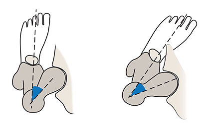
Top-view illustrations of excessive femoral anteversion
Left: Position of an anteverted femoral head with the foot facing straight forward. In this position, the femoral head subluxes out of the front of the hip joint.
Right: Most patients with excessive hip anteversion compensate by walking in-toed. This position keeps the femoral head within the socket, which minimizes pain.
Causes of femoral anteversion
The exact cause is unknown, however, femoral anteversion is congenital (present since birth) and develops while a child is in the womb. It appears to be related to the position of the baby while growing in the uterus. Since it often runs in families, it is believed that some people are genetically predisposed to the condition. This type of torsional deformity can also occur after trauma. After a femur fracture, a torsional malunion can occur, leading to same type of problems mentioned above.
Symptoms of femoral anteversion
- Signs and symptoms of femoral anteversion include:
- In-toeing, in which a person walks "pigeon-toed," with each foot pointed slightly toward the other.
- Bowlegs (also called bowed legs). Keeping the legs in this position often helps a patient maintain balance.
- Pain in the hips, knees and/or ankles.
- Snapping sound in the hip while walking.
Diagnosis of femoral anteversion
Generally, the doctor will review the patient's history, do a physical examination and observe the patient's gait (manner of walking) to look for signs of in-toeing. The physician may also order X-rays or a CT scan to look for any deformity. However, in certain cases, femoral anteversion can be difficult to detect. This is especially true in cases where the hip anteversion is combined with a separate rotational bone deformity, such as external tibial torsion – an outward rotation of the tibia (shinbone). This type of complex case is called "tetra-torsional malalignment,” which has sometimes also been called "miserable malalignment syndrome." It can be hard to diagnose because:
- The two opposite rotations of the femur and tibia leave the patient's feet to stay parallel during walking, This means the malalignment of the hips and knees may go unnoticed, even if the patient experiences pain or discomfort.
- X-rays, which are taken from the front, back or side, do not adequately display rotational deformities, which are on the axial plane and best viewed from above.
Treatment for femoral anteversion
While many children grow out of their femoral anteversion conditions, excessive anteversion may require surgical correction, as a procedure known as a femoral osteotomy. This surgery involves cutting and realigning the femur.
Patient story: Correction of miserable malalignment
Watch this video on limb rotational deformity correction with HSS patient Stephanie.
Articles related to femoral anteversion
Medically reviewed by S. Robert Rozbruch, MD
