Knee Surgery: High Tibial Osteotomy
Joint preservation surgery to repair damage to articular cartilage inflicted by knee osteoarthritis and malalignment
What is a high tibial osteotomy?
A high tibial osteotomy is a surgical procedure that realigns the knee joint. For some patients who have knee arthritis, this surgery can delay or prevent the need for a partial or total knee replacement by preserving damaged joint tissue.
What does this type of knee osteotomy treat?
A high tibial osteotomy knee surgery is performed primarily to treat medial, unicompartmental osteoarthritis of the knee (osteoarthritis that affects only the inner side of the knee) and/or a malalignment (incorrect angle) of bones that form the knee joint. Both conditions can cause the knee’s protective tissues to wear on one side more than the other in a repetitive cycle of damage. Similarly, it may also be indicated for people who have a meniscus injury in conjunction with a knock knee deformity. It is also sometimes used in the correction of bowlegs.
Osteoarthritis and malalignment of the knee
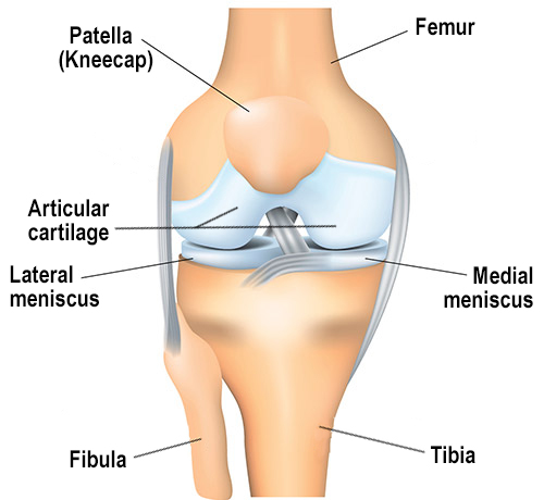
With each step you take, forces equal to 3 to 8 times your body weight travel between the femur (thighbone) and tibia (shin bone) in your knee. These forces are dampened by two menisci located on the inner and outer portion of the knee, and the ends of the bones are protected by articular cartilage.
Patients who have osteoarthritis of the knee experience a successive wearing on the menisci and articular cartilage, which may develop tears. The degeneration of these tissues limits the knee's ability to glide smoothly and can result in popping, catching, locking, clicking and pain.

In a condition called malalignment, unbalanced forces cause excessive pressure on either in the inner (medial) or outer (lateral) portion of the knee.
Who may benefit from a high tibial osteotomy?
Younger people with mild to moderate medial arthrosis (unicompartmental medial knee arthritis) and/or knee alignment problems may be able to delay or avoid knee replacement. A newly FDA-approved implantable shock absorber shows promise to provide an appropriate alternative to knee replacement or HTO for some patients. (Find a surgeon at HSS who performs HTO.)
High tibial osteotomy vs. knee replacement
When the knee joint damage is beyond repair, knee replacement surgery can correct this condition. But in certain patients, a high tibial osteotomy can realign the knee to take pressure off the damaged side by wedging open the upper portion of the tibia to reconfigure the knee joint. Weightbearing is then shifted away from the damaged or worn tissue and onto the healthier tissue. (Find a knee replacement surgeon at HSS to suit your specific condition, location and insurance.)
Because these benefits typically fade after 8 to 10 years, this type of osteotomy is generally considered to be a way to prolong the time before a knee replacement is necessary. This procedure is typically reserved for younger patients with pain resulting from instability and malalignment. An osteotomy may also be performed in conjunction with other joint preservation procedures in order to allow for cartilage repair tissue to grow without being subjected to excessive pressure.
How does a high tibial osteotomy work?
In an HTO, the surgeon first cuts the tibia bone close to the where it meets the femur to form the knee joint. Then, either a portion of bone is removed or an implant is inserted to realign the tibia and adjust the loadbearing forces on the knee when a patient stands, walks or runs. This reduces pressure on the medial compartment of the knee joint to relieve pain and improve mobility. The two basic techniques are called the closed wedge (or closing wedge) and the open wedge (or opening wedge) HTO.
Closed wedge high tibial osteotomy
After an initial osteotomy (cut through the bone) in the tibia close the knee joint, the orthopedic surgeon then makes a second cut to remove a wedge-shaped piece of bone from the tibia, which realigns it in relation to the femur.
Open wedge high tibial osteotomy
In this procedure, instead of a second cut to remove a wedge, the surgeon inserts a wedge-shaped implant into the tibia. This widens the tibia and reduces the pressure on the medial compartment of the knee. The open wedge HTO technique is usually performed in a person with more severe arthritis or who has a knock knee condition. In the latter case, it is often performed in conjunction with a distal femoral osteotomy.
Video: Animation of a high tibial osteotomy
High tibial osteotomy recovery
Recovery time for high tibial osteotomy can vary, depending on the severity of cartilage damage, in the case of knee arthritis, the general health of the patient, and other factors. In general, however, patients should expect 6 to 12 weeks of physical therapy with a complete recovery at around 3 to 6 months.
Authors
Attending Orthopedic Surgeon, Hospital for Special Surgery
Assistant Professor of Orthopedic Surgery, Weill Cornell Medical College
Attending Orthopedic Surgeon, Hospital for Special Surgery
Director of Research, Limb Lengthening and Complex Reconstruction Service, Hospital for Special Surgery
Attending Orthopedic Surgeon, Hospital for Special Surgery
Director, Limb Salvage and Amputation Reconstruction Center, Hospital for Special Surgery
Chief, Limb Lengthening and Complex Reconstruction Service, Hospital for Special Surgery
Director, Osseointegration Limb Replacement Center, Hospital for Special Surgery


