Bowlegs
Bowleg deformity is an incorrect alignment around the knee that can affect people of all ages. The condition is also known by various other common names and medical terms, including bow leg, bandy-leg, bowleg syndrome, bowed legs, varus deformity, genu varum, and tibia vara.
What are bowlegs?
Bowlegs refers to a condition in which a person’s legs appear bowed (bent outward) even when the ankles are together. It is normal in babies due to their position in the womb. But a child who still has bowlegs at about age three should be evaluated by orthopedic specialist.
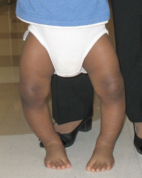
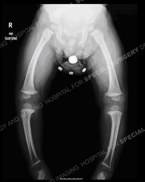
Photo and standing-alignment X-ray of a child with bowlegs.
Infants are often born bowlegged due to their folded positioning while in the mother’s womb. In typical growth patterns the child will outgrow this as they start to stand and walk. For this reason, up until the age of two, bowing of the legs is not unusual. In fact, there is a broad spectrum of what is considered normal. In most cases, the child’s legs begin to straighten once they start bearing weight on them while standing or walking (usually at between 12 to 18 months old). By age two to three, the leg angle typically reverses and begins to look more like knock knee, where the knees bend inward toward each other. After about six years of age, most children’s knees assume a straighter alignment that is considered normal.
A parent should consult a doctor to determine whether their child has the potential to develop a bowleg or knock knee deformity if, apart from the normal patterns discusses above:
- the angle of the child’s thighbone to shinbone falls outside of the normal range
- the direction the knees versus the direction the foot falls outside the normal pattern
- one leg is significantly more (or less) angled than the other
What are the symptoms of a bowleg deformity?
The most common symptom of a bowleg condition is that a person's knees do not touch while standing with their feet and ankles together. This causes a bowing of the legs that, if it continues beyond three years of age, suggests there is a bowleg deformity.
Other symptoms experienced by people with bowlegs include:
- knee pain
- hip pain
- reduced range of motion in hips
- difficulty walking or running
- knee instability
- unhappy feelings of appearance
Progressive knee arthritis is common in adults who were not diagnosed or treated for bowleg earlier in life. Adult patients who have had bowleg for many years overload the inside (medial compartment) and stretch the outside (lateral collateral ligament) leading to pain, instability, and arthritis. To prevent and delay the need for a knee replacement, an osteotomy should be performed to realign the knee.
What causes bowleg syndrome?
The many causes of bowleg syndrome range from illnesses such as Blount’s disease to improperly healed fractures, vitamin deficiencies and lead poisoning.
Illnesses and conditions that cause bowleggedness include:
- abnormal bone development (bone dysplasia)
- Blount’s disease (more information below)
- Paget’s disease (a metabolic disease impacting the way bones break down and rebuild)
- improperly healed fractures
- lead poisoning
- fluoride poisoning
- achondroplasia (the most common form of dwarfism)
- rickets (a bone-weakening disorder caused by a vitamin D deficiency)
- damage to growth plate
What is Blount’s disease?
Blount’s disease (also called tibia vara) is a growth disorder that affects the growth plates in the bones near the inside of the knee. Blount's slows down bone growth at these plates or halts growth there altogether. This leads to a bowlegged appearance and may also cause knee pain or instability.
The deformity occurs when the lateral (outer) side of the tibia continues to grow while the medial (inner) side of the bone does not. Blount’s disease may affect one or both legs. Most commonly, the growth deformity is found at the top of the tibia, which is the larger of the two bones in the lower leg. Blount’s disease may affect one or both legs.
Blount’s may be diagnosed in children aged two and up. Depending on the age at diagnosis, a child may have the infantile or adolescent type of this disease. Those with the infantile form of the disease are often early walkers. Obesity is frequently seen in children who have Blount’s, irrespective of the patient’s age. Adults who have not had treatment or inadequate treatment as children can present with large deformity and knee pain and degeneration.
How are bowlegs diagnosed?
Typically, a doctor will get the patient history, do a physical examination, and order a standing-alignment X-ray or EOS imaging of the leg bones from the hip to the ankle. Imaging helps the orthopedist determine the deformity’s location, magnitude and mechanical axis (where the bend occurs).
Do I need to get my bowlegs fixed?
If left untreated, people who are bowlegged may experience pain, increased deformity, knee instability and progressive knee degeneration (arthritis). Correction of the deformity leads to improved knee mechanics, better walking, less pain, and prevents the rapid progression of damage to the knee.
How are bowlegs treated?
Mild cases may be first carefully observed over time by a pediatric orthopedist. Bracing may be also tried to gradually correct the leg angles. When bracing is not enough, surgery may be recommended. Physical therapy also plays an important role, especially if surgery has been performed.
Surgical treatments
In a growing child, guided-growth, minimal-incision surgery may be used to encourage the limb to gradually grow straight.
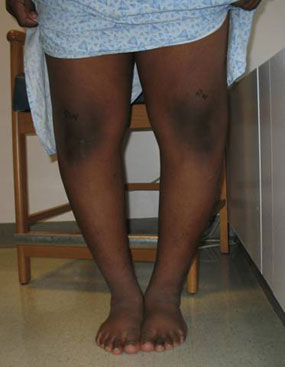
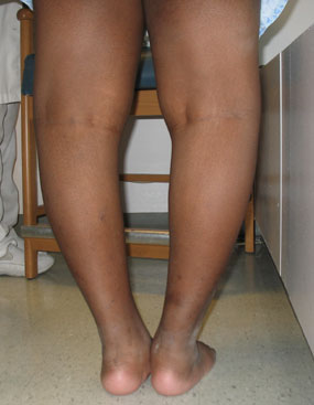
Photos from the front and back of a child with bowlegs due to Blount's disease.
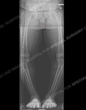
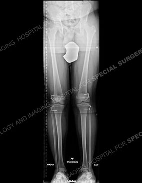
X-rays of the same child before (left) and after (right) a guided-growth surgical procedure. Note the plates used to fully correct the lower extremities in the knee area, highlighted in white.
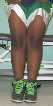
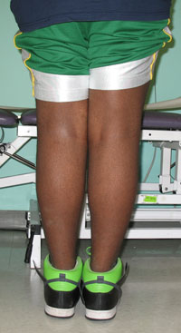
Photos of the child after surgery.
In skeletally mature adolescents and in adults, an osteotomy is the treatment to straighten the leg. X-rays are used to determine the location and magnitude of the deformity. In most cases, the tibia is treated, but there are situations when the femur or both femur and tibia are treated. When there is moderate deformity, the osteotomy is typically stabilized with internal fixation (a plate or rod inserted into the leg). When the malalignment is more severe, gradual realignment of the limb through the osteotomy is done with an external fixator. With external fixators, pins are inserted into the bone and protrude out of the body to attach to an external stabilizing structure. In some cases, the underlying bowleg condition causes one leg to be shorter than the other. This can also be corrected, using limb lengthening surgery.
Bowleg osteotomy surgery animation
This video shows an animation of a high tibial osteotomy (HTO) for bowleg correction in adults.
Articles about bowleg conditions
Learn more about when bowlegs are part of a child's normal growth pattern and when a bowleg condition falls outside of those patterns and should be assessed by a pediatric orthopedist.
Articles on treatments for bowlegs
Read more about various surgical treatments for bowleg conditions.
Articles on related issues
Learn about related conditions and how, at HSS, physical therapists and biomechanical engineers in the Leon Root, MD, Motion Analysis Lab assess bowleg patients after surgery to assess improvements in their walking ability.
Bowlegs Success Stories
In the news
Updated: 3/11/2021
Reviewed and updated by S. Robert Rozbruch, MD

