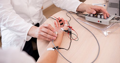
Electrodiagnostics
Electrodiagnostic testing is used to determine whether a patient has an injury or disorder of a nerve or muscle.

This type of testing is usually conducted to help diagnose patient who has experienced muscle pain or cramping, or sensations of weakness, numbness or tingling. It may be used in conjunction with a physical exam and radiological imaging, such ultrasound, magnetic resonance imaging (MRI) or magnetic resonance neurography (MRN).
Electrodiagnostics can also be used to monitor the recovery progress of some patients after they have been diagnosed. Conditions that can be diagnosed and/or monitored with the aid of electrodiagnostic testing include:
- Amyotrophic lateral sclerosis (ALS)
- Brachial plexus injuries
- Carpal tunnel syndrome
- Cubital tunnel syndrome (ulnar nerve entrapment)
- Herniated disc (“slipped disc”)
- Parsonage-Turner syndrome
- Radiculopathy
- Thoracic outlet syndrome
Electrodiagnostic testing actually involves two different tests:
- A nerve conduction study (NCS) – also known as a nerve conduction velocity (NCV) test
- An electromyography needle exam, also known as EMG testing.
For the nerve conduction study, the doctor stimulates a nerve with small electric shock by placing an electrode on the skin above the nerve. This test is done in two or more places along the nerve’s path. Data is recorded that shows the conduction velocity (the speed at which the electrical impulse travels through the nerve). If that speed is slower than typical readings, it suggests possible nerve damage.
For the EMG needle exam, the doctor lightly inserts very fine needles into a patient’s muscle (or several muscles). Connected to a computer, these needles record the electric impulses created by the body when the muscle is at rest and when the patient flexes the muscle. The physician then analyzes the recorded signals to determine whether there may be muscle or nerve damage. The EMG needles are so fine that there is rarely any bleeding during the test.
